Glutathione
Overview
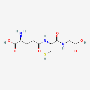
Glutathione is an endogenous tripeptide of glutamate, cysteine and glycine, linked by a γ-glutamyl bond and present mainly as reduced glutathione (GSH) and oxidised glutathione (GSSG). It occurs in some foods and is sold as a supplement, but it is not a vitamin, and intake alone does not determine functional status [1,2]. Availability depends on precursor supply, ATP-dependent synthesis, enzymatic recycling, cellular demand and the surrounding metabolic environment [1,2].
GSH supplies reducing capacity for glutathione peroxidases and selected glutathione S-transferase conjugations, and is regenerated from GSSG by NADPH-dependent glutathione reductase [1,2]. Oral glutathione can change biomarkers in some studies and not others; biomarker change is not by itself a clinical or brain outcome [3,4]. Mitochondria import glutathione rather than synthesising it de novo [5]. Synthesis as a BRS2 mechanism is covered on the glutathione-synthesis primary-mechanism page.
Key Compound Highlights
- Endogenous redox compound and tripeptide (γ-glutamyl-cysteinyl-glycine), not a vitamin and not adequately described as a general-purpose “antioxidant supplement” [1,2].
- GSH is the reduced form; GSSG is the oxidised disulfide. GSSG is not a synonym for GSH [1].
- Functional use includes peroxide reduction, selected electrophile conjugation and protein-thiol regulation; glutathione is an enzyme substrate or cosubstrate in those reactions, not an indiscriminate “detox” molecule [1,2].
- De novo synthesis is cytosolic and ATP-dependent; cysteine availability is often an important constraint, but the limiting factor varies by tissue and context [2].
- Mitochondria depend on glutathione imported from the cytosol [5].
- There is no dietary reference intake for glutathione. Food GSH and intracellular GSH are not equivalent [1,3,4].
Dietary Context
Food sources
Glutathione is measurable in some fresh foods, including uncooked meats, asparagus, avocado, spinach and other produce, but tabulated food values should not be treated as rankings of tissue glutathione support (Minich and Brown 2019). Content is variable; storage, processing and cooking can change measured amounts; digestion and first-pass handling mean food GSH is not equivalent to intracellular GSH [1]. This page therefore gives representative contexts rather than a food table with false precision.
Synergies
Dietary support is mainly about synthesis and recycling conditions, not about eating finished glutathione as if it were a vitamin. Distinguish:
- consuming glutathione already present in food;
- supplying amino-acid precursors through adequate protein, including cysteine and methionine within a balanced pattern, plus glycine and glutamate/glutamine context [2];
- supplying enzyme-related micronutrients, including selenium for glutathione peroxidase activity, riboflavin (FAD) for glutathione reductase, niacin-derived NADPH systems that support reduction capacity, and magnesium and cellular energy context for ATP-dependent synthesis [1,2];
- food bioactives that may influence Nrf2-related defence signalling, such as sulforaphane, without implying that one food raises brain glutathione [1];
- taking glutathione as a supplement, which is a separate intervention from precursor supply [3,4].
Methionine can contribute cysteine through transsulfuration, but dietary methionine is not a simple glutathione-boosting intervention [2]. Sulfur-containing foods may support precursor availability as part of overall protein quality. Do not infer that a named food raises brain glutathione unless direct human evidence shows that.
Supplement versus food
A food-first strategy normally concerns adequate precursor availability, dietary adequacy, selenium and riboflavin sufficiency, metabolic health, limiting avoidable oxidative exposures, and supporting synthesis and recycling rather than attempting to maximise glutathione indiscriminately. No conventional dietary reference intake exists, so this page does not set an intake target.
Supplement forms include standard oral reduced glutathione, liposomal and sublingual formulations, and S-acetyl glutathione. Intravenous or inhaled glutathione are medically supervised routes, not dietary equivalents. N-acetylcysteine (NAC) is a cysteine precursor with its own literature; it is not a glutathione formulation. Formulation, dose, study duration, baseline status and the compartment measured all affect findings [3,4].
An increase in circulating or cellular glutathione biomarkers does not by itself establish improvement in brain function or clinical outcomes.
Recipes
Foods
Biological Regulatory Systems
Compact BRS mapping for Nutritional Highlights. Deeper synthesis, recycling, genetics and measurement caveats are in Advanced Nutrition; condition-specific evidence is in Therapeutic Area Research. Mapping follows the evidence rather than forcing equal relevance across all six systems.
| Biological Regulatory System | Glutathione mapping | Evidence |
|---|---|---|
| Methylation & One-Carbon Metabolism (BRS2) | Strong relationship: methionine-cycle and transsulfuration can supply cysteine for glutathione synthesis. Substrate availability does not guarantee increased GSH synthesis. | [2] |
| Inflammation & Oxidative Stress (BRS3) | Primary functional relationship: peroxide reduction, redox buffering, selected electrophile conjugation and protein-thiol regulation. Oxidative demand and glutathione utilisation are reciprocal. | [1,2] |
| Mitochondrial Function & Bioenergetics (BRS4) | Strong cross-system relationship: mitochondria do not synthesise glutathione de novo and import it from the cytosol; ATP supports synthesis and NADPH supports GSSG reduction. | [2,5] |
| Neurotransmitter Regulation (BRS1) | Qualified: brain glutathione participates in neuronal and glial redox regulation. Oxidative conditions may influence receptor, membrane and synaptic environments. Glutathione is not a direct neurotransmitter treatment; total brain GSH by MRS is not a synaptic measure. | [1] |
| Gut–Brain Axis & Enteric Nervous System (BRS5) | Conditional: intestinal epithelial redox defence, microbial and dietary influences on precursor metabolism, and possible effects of gut inflammation on systemic oxidative demand. Probiotics or fermented foods have not been shown to raise brain glutathione reliably. | [1,2] |
| Metabolic & Neuroendocrine Regulation (BRS6) | Contextual: glycaemic dysregulation, inflammation and chronic stress may increase oxidative demand. Insulin and metabolic state can affect substrate handling. Composite allostatic load does not measure glutathione status. | [1,2] |
References
[1] Review of glutathione’s protective roles, measurement problems, and biosynthesis, including GSH/GSSG, glutathione peroxidases, glutathione S-transferases and recycling. Forman, Zhang and Rinna 2009. Glutathione: overview of its protective roles, measurement, and biosynthesis
[2] Review of glutathione synthesis and regulation: cytosolic glutamate-cysteine ligase and glutathione synthetase, ATP dependence, cysteine as a frequent constraint, and tissue differences. Lu 2013. Glutathione synthesis
[3] Six-month randomised trial of oral reduced glutathione in healthy adults: blood, erythrocyte, plasma, lymphocyte and buccal GSH rose at 250 or 1000 mg/day and returned toward baseline after washout. Secondary immune measures are not brain or clinical-outcome evidence. Richie et al. 2015. Randomized controlled trial of oral glutathione supplementation on body stores of glutathione
[4] Four-week oral glutathione trial (500 mg twice daily) in healthy adults: no change in GSH, GSSG or oxidative-stress biomarkers. Allen and Bradley 2011. Effects of oral glutathione supplementation on systemic oxidative stress biomarkers in human volunteers
[5] Mammalian glutathione is synthesised in the cytosol; SLC25A39 is necessary for mitochondrial glutathione import, with SLC25A40 as a related paralogue. Wang et al. 2021. SLC25A39 is necessary for mitochondrial glutathione import in mammalian cells
Advanced Nutrition
This panel is the biological overspill of the Highlights BRS mapping. It describes glutathione as a dynamic resource pool. Glutathione synthesis as a BRS2 primary mechanism is not reproduced here.
Synthesis pathway
De novo glutathione synthesis is cytosolic [2,5]. Glutamate and cysteine are joined by glutamate-cysteine ligase, a heterodimer of catalytic GCLC and modifier GCLM subunits, forming γ-glutamylcysteine. Glycine is then added by glutathione synthetase (GSS). Both steps require ATP [2]. GSH exerts feedback inhibition on glutamate-cysteine ligase, so synthesis is not a simple linear function of precursor intake [2].
Cysteine availability frequently exerts an important constraint, but the limiting factor varies by tissue and physiological context [2]. Plasma cysteine, intracellular cysteine and brain cysteine are not one pool. Cystine uptake through the cystine/glutamate antiporter (SLC7A11, system xc−) can supply intracellular cysteine in some cells; that transporter is not a universal brain-glutathione lever [1,2]. Methionine can contribute cysteine through transsulfuration via cystathionine β-synthase (CBS) and cystathionine γ-lyase (CTH), in competition with remethylation [2]. Substrate availability does not guarantee increased GSH synthesis.
Recycling and redox coupling
GSH is oxidised to GSSG during peroxide reduction and related thiol chemistry. Glutathione reductase (GSR) regenerates GSH using NADPH [1]. NADPH supply is linked to glucose-6-phosphate dehydrogenase (G6PD) and the pentose-phosphate pathway, and therefore to cellular glucose handling [1]. Glutathione peroxidases use GSH to reduce hydrogen peroxide and selected lipid hydroperoxides; selenium is required by the selenoprotein glutathione peroxidases [1]. Glutathione S-transferases conjugate GSH to selected electrophilic compounds. Glutaredoxin systems use GSH in protein-thiol regulation [1]. Export, extracellular breakdown and the γ-glutamyl cycle recover amino acids for resynthesis rather than acting as an unrestricted toxin-clearance pump [1].
The GSH:GSSG ratio is compartment-dependent and can be distorted by sampling and laboratory handling [1]. A single blood ratio is not a validated measure of brain redox status.
Related BRS3 context includes Nrf2–ARE antioxidant activation and antioxidant-network recycling. Related BRS4 context includes mitochondrial protection and redox integrity. The BRS2-to-BRS3 dependency describes one-carbon coupling to redox biology, not a proof that raising dietary glutathione corrects oxidative stress.
Resource-pool model
Synthesis, utilisation and recycling form a dynamic pool rather than a static nutrient store.
Inputs, synthesis, functional use, oxidation, and recycling or loss operate together. Lower measured GSH may represent increased use, impaired synthesis, redistribution, loss through conjugation and export, or methodological effects. Compensatory synthesis can maintain GSH under some conditions.
Cross-system cascade
The cascade is conditional and bidirectional. Compensatory synthesis may maintain GSH under some conditions. Lower measured GSH is not proof of a primary glutathione-deficiency disease, and the cascade is not proof that supplementation will correct the originating condition.
Genetics and individual variation
Pathway genes of biochemical interest include GCLC, GCLM, GSS, GSR, SLC7A11, CBS, CTH, glutathione peroxidase and glutathione S-transferase families, and G6PD where NADPH supply is relevant [1,2]. Rare pathogenic enzyme deficiencies can cause clinically recognised glutathione-synthesis disorders [2]. Common functional polymorphisms and association studies are not equivalent to those rare diseases. Direct-to-consumer SNP interpretations are not clinically actionable here.
Common GST deletions or a single SNP are not proof of “poor detoxification.” This page does not include a consumer SNP interpretation table.
Measurement
Whole blood, erythrocyte, plasma and cellular glutathione are different compartments [1]. GSH and GSSG must be distinguished, and assay handling (oxidation after collection, delayed processing, haemolysis) can distort ratios [1]. Tissue specificity matters: hepatic glutathione is not erythrocyte glutathione, and neither is brain glutathione. Magnetic resonance spectroscopy can estimate total brain GSH in some regions; that signal is not interchangeable with peripheral GSH, synaptic GSH, mitochondrial GSH or the GSH:GSSG ratio [1]. Peripheral findings cannot automatically be translated into brain glutathione status.
Supplement evidence and safety
Marketing sometimes calls glutathione the “master antioxidant.” That phrase is not used here as a scientific classification. More glutathione is not always better.
| Form or strategy | What it provides | Evidence considerations |
|---|---|---|
| Oral reduced glutathione | Finished GSH | Human biomarker findings are mixed and formulation-dependent. A six-month trial reported increased body-store biomarkers [3]; a four-week trial did not [4]. An older oral-availability study did not find a rise in circulating GSH after a large single oral dose (Witschi et al. 1992). |
| Liposomal glutathione | Encapsulated GSH | A small one-month study (n=12) reported biomarker changes (Sinha et al. 2018). Independent replication and clinical outcomes remain limited. |
| Sublingual glutathione | Alternative delivery route | Limited comparative evidence. |
| S-acetyl glutathione | Modified derivative | Human bioavailability and outcome evidence remain insufficient for bioavailability claims. |
| NAC | Cysteine precursor | Separate substance with its own efficacy and safety literature. NAC trials are not glutathione-supplementation trials. |
| Glycine plus NAC | Precursor combination | Emerging clinical research, including precursor work in older adults (Sekhar et al. 2011). Do not generalise from older or metabolically impaired populations. |
| Whey or protein interventions | Cysteine-containing food matrix | Effects depend on composition, digestion and baseline status. |
| Intravenous glutathione | Medical administration | Not equivalent to dietary supplementation; requires clinical oversight. Not recommended here. |
This table does not rank products and does not recommend doses.
Safety considerations include common gastrointestinal effects with oral products; bronchospasm concerns with inhaled glutathione in people with asthma (Marrades et al. 1997); medication and clinical-context cautions; uncertain long-term effects of unnecessary high-dose use; and supplement quality and formulation variability. Use in pregnancy, childhood and chronic disease requires appropriate professional assessment. Intravenous glutathione is not a food-first intervention and is not recommended here.
References
- Forman, Zhang and Rinna 2009. Protective roles, measurement and biosynthesis.
- Lu 2013. Synthesis, GCLC/GCLM, GSS, cysteine constraint and regulation.
- Meister and Anderson 1983. Foundational biochemistry of glutathione and the γ-glutamyl cycle.
- Wang et al. 2021. Cytosolic synthesis and mitochondrial import via SLC25A39.
- Richie et al. 2015. Longer-term oral glutathione and body-store biomarkers.
- Allen and Bradley 2011. Four-week oral glutathione without biomarker change.
- Witschi et al. 1992. Older oral systemic-availability study.
- Sinha et al. 2018. Small liposomal glutathione biomarker study.
- Sekhar et al. 2011. Precursor (cysteine and glycine) support of glutathione synthesis in older adults.
- Zalachoras et al. 2022. Accumbal GSH, MRS and motivated effort; human association plus mechanistic animal work, not an ADHD or supplement trial.
- Marrades et al. 1997. Nebulised glutathione induced bronchoconstriction in mild asthma.
Therapeutic Area Research
This panel collects condition-specific evidence. It is not a treatment protocol. Observational biomarker findings, mechanistic plausibility and intervention evidence are listed separately. NAC trials are not glutathione-supplementation trials. Therapeutic-area pages are not currently published; the names below are labels, not links.
Condition evidence matrix
| Therapeutic area | Observational evidence | Mechanistic relevance | Intervention evidence | Framework status |
|---|---|---|---|---|
| ADHD | Oxidative-stress findings are heterogeneous; antioxidant status was not significantly different in a meta-analysis, and oxidative-stress association lost significance after clustering correction (1). Peripheral GSH/GSSG reports exist in small cohorts (2). Brain GSH deficiency is not established. | Immune-redox and bioenergetic plausibility through peroxide reduction and mitochondrial import | No controlled oral-glutathione efficacy trials in ADHD. NAC or pine-bark extract trials are not glutathione trials (2). | Mechanistic and hypothesis-generating |
| Autism spectrum disorder | Observational redox and glutathione abnormalities reported, including impaired methylation-capacity biomarkers in children (3). | Shared transsulfuration and redox biology | Precursor (NAC) pilot evidence is not glutathione-formulation evidence (4). Findings are not transferable to ADHD. | Investigational |
| Depression and other psychiatric conditions | Mixed peripheral markers; limited MRS work. Accumbal GSH has been associated with motivated effort in a human MRS study that is not a depression trial (5). | Neuronal and glial redox regulation | No established glutathione treatment for depression or ADHD-related mood symptoms. | Associative / hypothesis-generating |
| Neurodegenerative disorders | Post-mortem glutathione reduction in Parkinson’s disease substantia nigra (6). Alzheimer’s and related conditions have been investigated with mixed biomarker findings. | Mitochondrial redox protection and peroxide handling | Small or route-specific intervention studies exist; none establish oral glutathione as a disease treatment. | Mechanistically relevant |
| Metabolic and hepatic contexts | Liver is a major glutathione-synthesis organ (7, 8). | Electrophile conjugation and peroxide reduction in hepatocytes | Clinical toxicology and metabolic research are not transferable to brain-health supplementation or “liver detox” marketing. | Established biochemistry; not a brain-treatment claim |
ADHD
Oxidative-stress findings in ADHD are heterogeneous. A meta-analysis of oxidative-stress and antioxidant measures found that antioxidant status was not significantly different in ADHD, and the oxidative-stress association lost statistical significance after correction for clustering of multiple measures from the same studies (1). Altered oxidative biomarkers do not demonstrate a primary glutathione deficiency. Peripheral GSH or GSSG findings cannot automatically be translated into brain GSH status.
A small randomised pine-bark extract study reported GSH/GSSG changes in children with ADHD; that intervention was not glutathione (2). Direct glutathione-supplementation trials in ADHD were not identified. NAC evidence, where it exists in other conditions, is not equivalent to glutathione evidence.
Glutathione is biologically relevant to the immune-redox and bioenergetic systems implicated in ADHD, but current evidence does not establish a general glutathione-deficiency subtype of ADHD or support glutathione supplementation as an established ADHD treatment. Its role within the framework is therefore mechanistic and hypothesis-generating unless direct clinical evidence becomes available.
Autism spectrum disorder
Observational studies have reported redox and glutathione-related abnormalities in some autism-spectrum cohorts, including metabolic biomarkers of oxidative stress and impaired methylation capacity (3). Precursor interventions such as NAC have been studied in small randomised pilots with behavioural outcomes (4). Those studies are not glutathione-formulation trials, and autism findings should not be transferred to ADHD. Symptom outcomes, study size and replication remain limited.
Depression and other psychiatric conditions
Evidence with adequate human support is limited. Peripheral glutathione markers and occasional MRS findings should not be collapsed into a treatment claim. A human MRS study linked nucleus-accumbens GSH to motivated effort; complementary rodent work used synthesis inhibition and NAC (5). That work is not a depression or ADHD supplementation trial and does not establish oral glutathione as a psychiatric treatment.
Neurodegenerative disorders
Parkinson’s disease has a long-standing post-mortem finding of reduced glutathione in the substantia nigra (6). Alzheimer’s disease and other neurodegenerative conditions have been investigated through redox and mitochondrial hypotheses. Mechanistic rationale, biomarker findings and controlled intervention evidence remain separate. No oral glutathione protocol is established as a neurodegenerative treatment on this page.
Metabolic and hepatic contexts
The liver is a central site of glutathione synthesis and of glutathione-dependent conjugation of selected electrophilic compounds (7, 8). That biochemistry is relevant to metabolic and toxicological research. It does not license liver “detox” marketing, nor does hepatic glutathione biology automatically transfer to brain-health supplementation.
Evidence conclusion
- Endogenous synthesis and redox functions: established
- Role as substrate for glutathione-dependent enzymes: established
- Nutritional dependence of synthesis and recycling: established in principle
- Direct equivalence between food glutathione and intracellular GSH: not established
- Oral supplementation increasing biomarkers: formulation- and study-dependent
- Biomarker improvement establishing clinical benefit: not established generally
- General glutathione deficiency in ADHD: not established
- Glutathione supplementation as ADHD treatment: not established
- Precision use based on common SNPs: not established
References
[1] ADHD matrix row. Meta-analysis of oxidative-stress and antioxidant measures in ADHD; antioxidant status was not significantly different, and oxidative-stress association lost significance after clustering correction. Joseph et al. 2015. Oxidative Stress and ADHD: A Meta-Analysis
[2] ADHD matrix row. GSH/GSSG changes in children with ADHD during a pine-bark extract trial, not a glutathione-supplementation trial. Dvořáková et al. 2006. The effect of polyphenolic extract from pine bark, Pycnogenol, on the level of glutathione in children suffering from attention deficit hyperactivity disorder (ADHD)
[3] Autism matrix row. Observational metabolic biomarkers of oxidative stress and impaired methylation capacity in children with autism. James et al. 2004. Metabolic biomarkers of increased oxidative stress and impaired methylation capacity in children with autism
[4] Autism matrix row. Randomised pilot of oral N-acetylcysteine in children with autism; precursor evidence, not glutathione-formulation evidence. Hardan et al. 2012. A randomized controlled pilot trial of oral N-acetylcysteine in children with autism
[5] Depression and other psychiatric conditions matrix row. Human MRS association of accumbal GSH with motivated effort, plus rodent mechanistic work; not a psychiatric glutathione trial. Zalachoras et al. 2022. Glutathione in the nucleus accumbens regulates motivation to exert reward-incentivized effort
[6] Neurodegeneration matrix row. Post-mortem glutathione reduction in Parkinson’s disease substantia nigra. Sian et al. 1994. Alterations in glutathione levels in Parkinson's disease and other neurodegenerative disorders affecting basal ganglia
[7] Metabolic and hepatic matrix row. Review of glutathione synthesis and hepatic regulation. Lu 2013. Glutathione synthesis
[8] Metabolic and hepatic matrix row. Review of glutathione biochemistry, including conjugation and redox functions. Forman, Zhang and Rinna 2009. Glutathione: overview of its protective roles, measurement, and biosynthesis