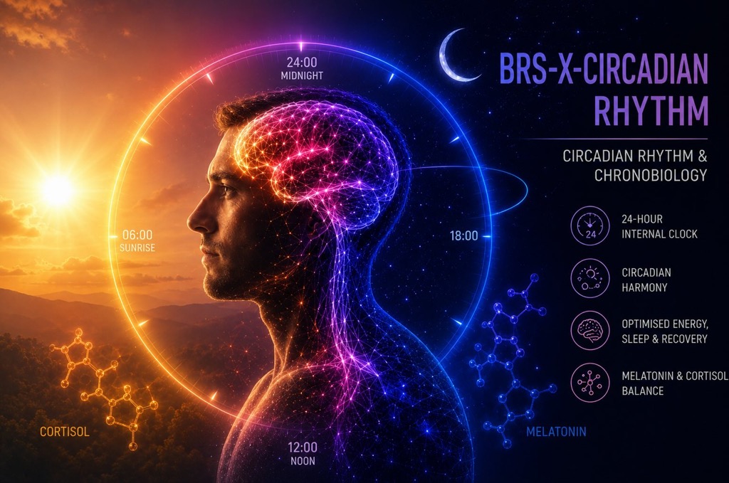
BRS-X(Circadian-FM1-PM2) - Feeding-Time Circadian Synchronisation
(Meal Timing and Peripheral Clocks)
1. Mission & Overview
Mission
Coordinate peripheral metabolic clock timing through feeding cues.
Intervention Dominance: Lifestyle-Dominant — System Optimisation Practices
Feeding-driven peripheral timing is distinct from metabolic benefit and central-clock resetting. Principal route selection identifies the mechanism-specific intervention class, not a universal optimal schedule or superiority across all health outcomes.
- Defined meal-timing schedule
Overview
Feeding time is a cue for peripheral metabolic clocks, helping organise when tissues process incoming nutrients. Changing meal timing can shift these rhythms without shifting the measured central-clock markers. Human evidence distinguishes the timing of blood glucose from the timing of a clock gene in adipose tissue. The practical route is a defined meal schedule; changing timing is not automatically evidence of better alignment or health. Wehrens et al. (2017) [1]; Stokkan et al. (2001) [4]
- Implementation Notes: Use the defined meal-schedule evidence below to understand which rhythms can respond. It does not prescribe delaying meals or a universal fasting window. Wehrens et al. (2017) [1]
- Biological Relevance: Peripheral timing can change while measured central markers remain stable. Cortisol regulation remains with BRS6 PM4. Wehrens et al. (2017) [1]
2. Primary Biological Effects
- Glucose rhythm delayed by 5.69 ± 1.29 hours and adipose PER2 by 0.97 ± 0.29 hours after a five-hour meal delay. Wehrens et al. (2017) [1]
- No significant shift of measured melatonin or cortisol timing in that protocol. Wehrens et al. (2017) [1]
3. Levers
No Direct or Derived Dietary Requirement is currently established for synchronising peripheral clocks with feeding time.
No evidence-supported cofactors or substrates are currently established for this mechanism.
No mapping established.
No evidence-qualified lifestyle relationship is projected.
4. Mechanistic Basis
Summary
Feeding cues can reset peripheral rhythms independently of the light-linked central pacemaker. Human glucose and adipose clock-gene responses differ in magnitude; animal insulin/IGF-1 experiments support a mechanistic route. Wehrens et al. (2017) [1]; Crosby et al. (2019) [6]
Mechanism boundary: Light-driven central entrainment remains with PM1. Glucose concentration, weight change and inflammatory outcomes do not independently demonstrate clock resetting. This PM does not own cortisol synthesis or microbial substrate-processing capacity.
4.1 Scientific Findings
Summary
Human meal scheduling shifted glucose timing much more than adipose PER2 timing. Animal studies establish independent peripheral resetting and a feeding-hormone-to-PERIOD pathway. These findings support a timing mechanism; they do not show that every phase shift improves alignment, or that metabolic protection alone demonstrates entrainment.
In ten healthy men, a five-hour meal delay shifted glucose rhythm timing by 5.69 ± 1.29 hours and adipose PER2 by 0.97 ± 0.29 hours, without significant changes in measured melatonin or cortisol timing.
What this means
Peripheral metabolic timing can move while the central melatonin and cortisol signals stay put. A metabolic benefit is not that clock result.
Evidence confidence: Not yet scored
Finding ID: PM2-F1
Finding Statement: In ten healthy men, a five-hour meal delay shifted glucose rhythm timing by 5.69 ± 1.29 hours and adipose PER2 by 0.97 ± 0.29 hours, without significant changes in measured melatonin or cortisol timing.
Synthesised Evidence Confidence: Not yet scored
Synthesis: A 13-day ordered laboratory protocol used early meals then six days of late meals. Rhythms were assessed during 37-hour constant routines with hourly snacks. The glucose rhythm shifted much more than adipose PER2; these endpoints are not interchangeable and do not demonstrate improved whole-system alignment.
Synthesis Limitations: The sample is ten young men on an ordered laboratory schedule. It does not establish a universal eating window.
Evidence Considered:
- Study
- Ten healthy men; meals five hours apart, starting 0.5 versus 5.5 hours after waking; 37-hour constant-routine rhythm assessments.
- Population
- Ten healthy men.
- Result
- In ten healthy men, a five-hour meal delay shifted glucose rhythm timing by 5.69 ± 1.29 hours and adipose PER2 by 0.97 ± 0.29 hours, without significant changes in measured melatonin or cortisol timing.
- Effect / Magnitude
- Glucose phase +5.69 ± 1.29 h; adipose PER2 +0.97 ± 0.29 h; these are measured responses, not the five-hour exposure dose.
- Evidence Summary
- Meal timing changed peripheral metabolic and adipose-clock readouts without moving the measured central hormonal timing.
- Limitations
- Small ordered rather than randomised sequence; peripheral clock-gene sampling, not direct SCN recording. No optimum eating window or clinical benefit established.
- Evidence Source
- Inherited repository evidence
- Reference
- [1]
Connected / Supportive Evidence:
- Hatori et al. (2012) [2] — Animal DataWhy relevant: Time-restricted feeding without a calorie reduction prevented metabolic disease in mice on a high-fat diet.Why excluded from the primary synthesis: Metabolic protection is not a measurement of peripheral clock phase in humans.
- Garaulet and Gómez-Abellán (2014) [3] — ReviewWhy relevant: The review associates the timing of food intake with obesity.Why excluded from the primary synthesis: An association with obesity does not establish that a peripheral clock moved.
Animal feeding manipulations shift peripheral clock rhythms while central-clock phase remains linked to the light cycle.
What this means
Stokkan and Damiola distinguish food-driven peripheral resetting from light-linked central timing. Crosby identifies insulin/IGF-1 signalling and PERIOD induction as a mechanistic link. This supports a feeding-to-clock chain, not a general recommendation to increase insulin or nutrient intake.
Evidence confidence: Not yet scored
Finding ID: PM2-F2
Finding Statement: Animal feeding manipulations shift peripheral clock rhythms while central-clock phase remains linked to the light cycle.
Synthesised Evidence Confidence: Not yet scored
Synthesis: Stokkan and Damiola distinguish food-driven peripheral resetting from light-linked central timing. Crosby identifies insulin/IGF-1 signalling and PERIOD induction as a mechanistic link. This supports a feeding-to-clock chain, not a general recommendation to increase insulin or nutrient intake.
Synthesis Limitations: Animal and cellular pathways support the mechanism; their tissue responses and effect sizes cannot be assigned to every human organ or feeding protocol.
Evidence Considered:
- Study
- Transgenic rat tissue rhythms under restricted feeding.
- Population
- Transgenic rat tissue rhythms under restricted feeding.
- Result
- Liver rhythm shifted by approximately ten hours within two days while SCN rhythmicity stayed phase-locked to the light–dark cycle.
- Effect / Magnitude
- Liver rhythm shifted by approximately ten hours within two days while SCN rhythmicity stayed phase-locked to the light–dark cycle.
- Evidence Summary
- Liver rhythm shifted by approximately ten hours within two days while SCN rhythmicity stayed phase-locked to the light–dark cycle.
- Limitations
- Animal liver entrainment, not a human optimum meal schedule.
- Evidence Source
- Bounded external search
- Reference
- [4]
- Study
- Mouse daytime/night-time restricted feeding with peripheral and SCN clock-gene measurements.
- Population
- Mouse daytime/night-time restricted feeding with peripheral and SCN clock-gene measurements.
- Result
- Peripheral gene-expression phase shifted by up to twelve hours while SCN phase remained unaffected; adaptation differed across tissues.
- Effect / Magnitude
- Peripheral gene-expression phase shifted by up to twelve hours while SCN phase remained unaffected; adaptation differed across tissues.
- Evidence Summary
- Peripheral gene-expression phase shifted by up to twelve hours while SCN phase remained unaffected; adaptation differed across tissues.
- Limitations
- Peripheral uncoupling can occur; a shift is not proof of beneficial alignment.
- Evidence Source
- Bounded external search
- Reference
- [5]
- Study
- Cellular and mouse insulin/IGF-1 signalling manipulations.
- Population
- Cellular and mouse insulin/IGF-1 signalling manipulations.
- Result
- Insulin/IGF-1 induced PERIOD proteins and reset rhythms; mistimed signalling disrupted circadian organisation.
- Effect / Magnitude
- Insulin/IGF-1 induced PERIOD proteins and reset rhythms; mistimed signalling disrupted circadian organisation.
- Evidence Summary
- Insulin/IGF-1 induced PERIOD proteins and reset rhythms; mistimed signalling disrupted circadian organisation.
- Limitations
- Animal/cellular mechanism, not proof that a particular human diet optimises clocks.
- Evidence Source
- Bounded external search
- Reference
- [6]
5. BRS Pathways and Connections
5.1 BRS Pathways
- None listed
5.2 Cross-BRS Mechanism Relationships
Primary Mechanisms in other Biological Regulatory Systems that directly interact with, constrain or support this mechanism.
- BRS6-FM2-PM4 — Cortisol Rhythm Regulation — cortisol timing did not move with the meal delay; cortisol output is a different job; see PM4
- BRS6-FM1-PM1 — Glucose Appearance Kinetics — a shift in glucose timing is not the same job as glucose appearance; see BRS6-FM1-PM1
- BRS5-FM2-PM4 — Microbial Substrate-Processing Selection & Adaptation — feeding schedules are an exposure context; those substrate studies do not establish clock synchronisation
5.3 Local BRS Mechanism Relationships
Related Primary Mechanisms within the same Biological Regulatory System that collectively support the integrated biological function.
- BRS-X(Circadian-FM1-PM1) — Light–Dark Circadian Entrainment — retinal light changes a central melatonin signal; see PM1
7. Phenome Connections
These mappings are translational relationships, not single-mechanism outcome claims. Phenomes are emergent functional patterns supported by multiple interacting PMs across the BRAIN Framework.
Each Phenome relationship has two independent ratings. They are not combined or averaged.
Biology → Phenome Relationship Strength
How direct and important is this biological mechanism expected to be to the Phenome within the BRAIN model?
Evidence Confidence
How strongly does the adjudicated evidence support this particular biology → Phenome relationship?
No direct functional outcome relationship currently mapped.
8. References
- [1] Wehrens et al. (2017) — Meal Timing Regulates the Human Circadian System
- [2] Hatori et al. (2012) — Time-restricted feeding without reducing caloric intake prevents metabolic diseases in mice fed a high-fat diet
- [3] Garaulet & Gómez-Abellán (2014) — Timing of food intake and obesity: a novel association.
- [4] Stokkan et al. (2001) — Entrainment of the circadian clock in the liver by feeding
- [5] Damiola et al. (2000) — Restricted feeding uncouples circadian oscillators in peripheral tissues from the central pacemaker in the suprachiasmatic nucleus
- [6] Crosby et al. (2019) — Insulin/IGF-1 Drives PERIOD Synthesis to Entrain Circadian Rhythms with Feeding Time